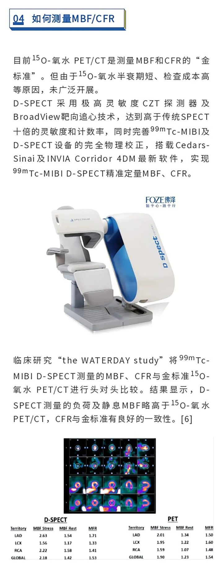
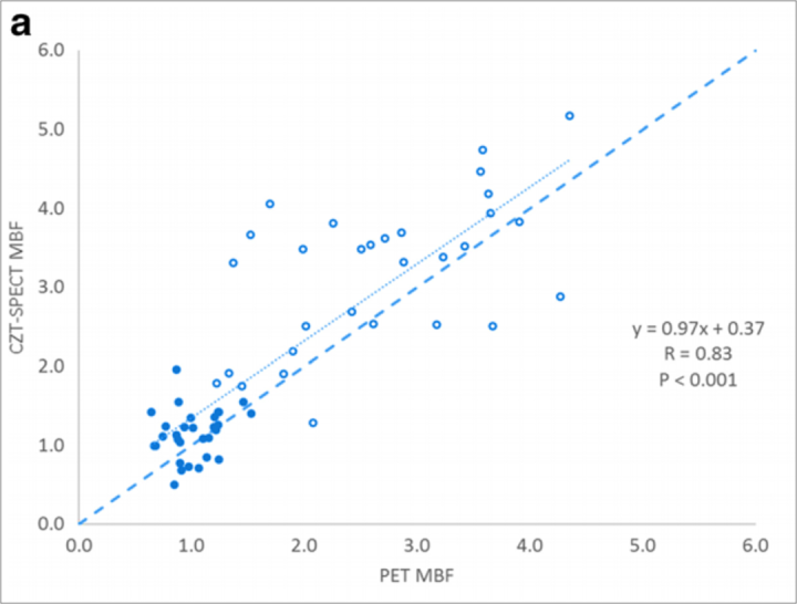
 D-SPECT 与 PET测量心肌血流一致性
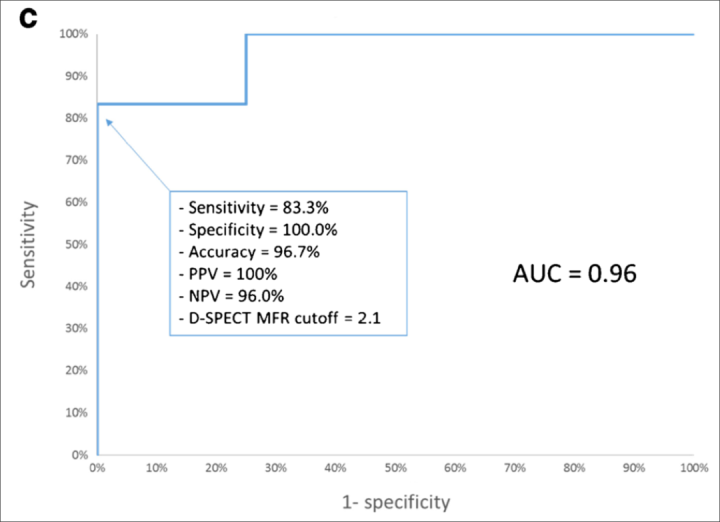 D-SPECT CFR灵敏度、特异性高,诊断符合率高达96.7%
1. Gould KL. Does coronary flow trump coronary anatomy? JACC Cardiovasc Imaging 2009;2(8):1009-23.
2. 冠状动脉血流储备分数临床应用专家共识专家组.冠状动脉血流储备分数临床应用专家共识[J] . 中华心血管病杂志,2016,44(4): 292-297.
3. Manabe O, Naya M, Tamaki N. Feasibility of PET for the management of coronary artery disease: Comparison between CFR and FFR. Journal of cardiology. 2017 Aug 1;70(2):135-40.
4. van de Hoef TP, van Lavieren MA, Damman P, Delewi R, Piek MA, ChamuleauSA, Voskuil M, Henriques JP, Koch KT, de Winter RJ, Spaan JA, Siebes M, TijssenJG, Meuwissen M, Piek JJ. Physiological basis and long-term clinical outcome ofdiscordance between fractional flow reserve and coronary flflow velocity reserve in coronary stenoses of intermediate severity. Circ Cardiovasc Interv2014;7(3):301–11.
5. Patel KK, Spertus JA, Chan PS, et al. Myocardial blood flow reserve assessed by positron emission tomography myocardial perfusion imaging identifies patients with a survival benefit from early revascularization. European heart journal 2020; 41(6): 759-68.
6. Agostini D, Roule V, Nganoa C, et al. First validation of myocardial flow reserve assessed by dynamic (99m)Tc-sestamibi CZT-SPECT camera: head to head comparison with (15)O-water PET and fractional flow reserve in patients with suspected coronar artery disease. The WATERDAY study. Eur J Nucl Med Mol Imaging. 2018;45(7):1079-90.


